As clinicians at a university teaching hospital, we are privy to some unique cases. Often, they are referred by a veterinarian who is struggling to make a diagnosis or has one in mind that needs a deeper dive into diagnostics for a confirmation. Occasionally, we are presented with what seems like a perfectly normal case that turns out to be a once-in-a-career diagnosis: a “unicorn.”
A seemingly normal 8-year-old red roan Quarter Horse mare arrived at the Equine Reproduction Laboratory, whose owner wanted her to become pregnant and carry her own foal. Reported to be a true “maiden mare” (never been bred, never had a foal), her reproductive history was very short. She arrived in late January with a heavy winter coat, a product of the cold, Colorado weather. Despite that, initial ultrasound examination identified the presence of a corpus luteum on one ovary, indicating that she ovulated recently and would likely continue to cycle. However, there was something quite different about her rectal palpation findings. Instead of detecting two distinct uterine horns, it appeared the left uterine horn was absent, the uterus being manually palpated as a simple ligamentous structure. Transrectal ultrasonography recognized the same abnormal finding, which was exceptionally puzzling due to the presence of a functional left ovary, presumably unattached to any uterine structure.
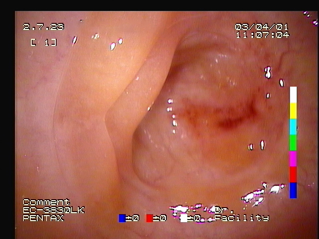
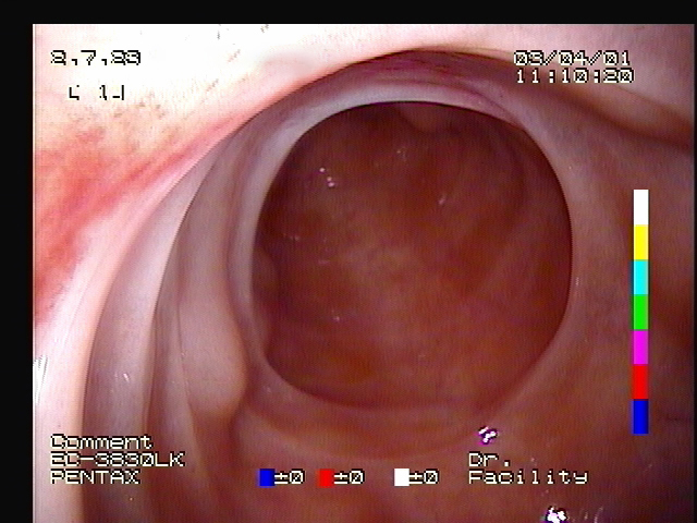
These findings were discussed with the owner with a presumptive diagnosis of uterus unicornis, or a single uterine horn. Several options were reviewed, but a confirmation needed to be made through a visual inspection. A hysteroscope is a tool used in medical practice with a video scope and light source for viewing the internal anatomy of the uterus through the cervix. In this case, it was used to rule in or rule out the physical presence of a left uterine horn. The mare was placed in exam stocks and given a standing sedative agent to provide a quiet and calm atmosphere to perform the hysteroscopic procedure. Routine insufflation with air allowed for visual inspection of her uterus, which confirmed a single (right) uterine horn without the presence of the left uterine horn. Additionally, there was no evidence of an opening or scar tissue to suggest that the horn had been surgically removed. All features associated with the right uterine horn were confirmed as normal.
Uteris unicornis in mares
Uterus unicornis is a rare congenital disorder reported throughout veterinary literature in a variety of domestic species, including mares. These cases most commonly present as decreased fertility, likely attributed to ovulations occurring upon the ovary unattached to the absent uterine horn. Pregnancies have been reported, but due to the unique placentation of the mare and a decreased surface area associated with this condition, fetal complications or reduced growth are concerns if a pregnancy is allowed to occur. No chromosomal abnormality has been identified and therefore no inheritance has been implicated. As a disorder affecting the developmental component of the ductal system during gestation, other factors such as environmental conditions could be implicated.
Following the confirmation of this unique anatomical anomaly, options were once again discussed with the owner to still reach the ultimate goal of obtaining a single offspring from this mare for the breeding season. The first decision made was to surgically remove the left ovary, as it was unattached and would not be able to provide a viable oocyte (egg) for fertilization. By removing this ovary, she would only be able to ovulate from the right ovary which would provide a more consistent opportunity for fertilization to occur. It was also determined that this mare’s unique uterine anatomy would not provide an adequate amount of space for a developing foal and could pose risk to the pregnancy and/or the mare’s health if allowed to carry her own pregnancy. Therefore, an embryo transfer technique would provide the best option in this scenario to reach the owner’s goal.
Embryo transfer
Embryo collection and transfer procedures are an excellent option for owners to pursue with their own “unicorns” and not necessarily reserved for unique anatomical anomalies. Many owners who have a mare with a busy show schedule, want to achieve multiple pregnancies each year, or simply do not wish to allow their mare to carry a pregnancy elect to utilize this procedure each year. To perform this procedure, the reproductive cyclicity of the owner’s mare (donor) is managed by a team of veterinarians to determine the optimal time for artificial insemination. Either fresh, cooled-shipped, or frozen semen can be used to inseminate the mare, but once ovulation occurs the team waits a little over a week before attempting to collect a tiny embryo. This part of the procedure involves infusion of a specialized media into her uterus and allowing it to flow back out through a filtration device in hopes of capturing that tiny embryo. If successfully collected, the tiny embryo is gently transferred into the uterus of a surrogate (recipient) mare who will nourish and carry the pregnancy to term.
For this “unicorn” mare, she was allowed to appropriately recover from the removal of her left ovary before continuing reproductive monitoring. Interestingly, the laparoscopic removal of her ovary allowed surgeons to again confirm the absence of a tubular uterine horn replaced by a thin membranous band. Now as the only ovary, the right side was examined with transrectal ultrasonography until impending ovulation whereupon cooled-shipped semen arrived from Texas and the mare was artificially inseminated. Roughly a week later through the embryo collection procedure, an embryo was successfully collected and transferred into a selected recipient mare. To everyone’s delight, this recipient mare was confirmed pregnant a few days later! The growing pregnancy was examined routinely to determine that important milestones were being met through early gestation. When ready, both recipient mare and the unicorn were picked up to return home with a grateful owner.
As described, this congenital abnormality is rare, but even more rare is the opportunity to achieve a pregnancy from a mare with this diagnosis. Through the collaborative efforts of surgeons and theriogenologists at Colorado State University, this owner will hopefully have a healthy foal next year!