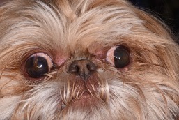
Components of brachycephalic ocular syndrome include:
- Shallow orbits resulting in protrusion of the eyes and inability to close the eyelids fully (particularly while sleeping)
- Macropalpebral fissure (excessively wide opening of the eyelids) resulting in over-exposure of the cornea (transparent, domed surface) and white of the eye
- Abnormal eyelid conformation resulting in rubbing of facial hairs on the surface of the eye
These abnormalities can predispose patients to potentially serious ocular disease including pigmentary keratitis (accumulation of pigment on the clear surface of the eye), corneal ulceration (wound on the surface of the eye), dry eye (due to exposure), and potential to experience traumatic proptosis (protrusion of the eye with the eyelids stuck behind the globe) due to minor trauma.
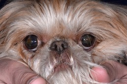
A medial canthoplasty is a surgical procedure used to correct over-exposure of the eyeball and provide additional protection to the eye due to brachycephalic ocular syndrome. The procedure is most commonly recommended for short-nosed dogs such as pugs, pekingese, and shih tzus with brachycephalic ocular syndrome.
The medial canthoplasty procedure aims to decrease the excessively wide opening in the eyelids while correcting abnormal hairs contacting the cornea and improving your pet’s ability to close their eyes fully.