Early accurate diagnosis of pregnancy status, detection and management of twins and identification of pregnancy loss are all important for optimal reproductive management of mares.
Transrectal Ultrasound Pregnancy Examination
The recommended method for initial pregnancy examination is transrectal ultrasonography performed 14 to 16 days after ovulation, or earlier if needed. This protocol allows for accurate diagnosis of pregnancy status, early detection and management of twins, evaluation of the corpus luteum, and assessment of the presence or absence of uterine edema and uterine fluid. A thorough, systematic ultrasound examination of the entire reproductive tract should be performed. The entire uterus should be scanned in one continuous motion in order to avoid missing any section of the uterus. The most common locations for a single embryo or a twin embryo to be missed are the caudal uterine body adjacent to the cervix or the tip of a uterine horn.
An embryonic vesicle may be detected as early as day 10 or 11 after ovulation. Routine initial pregnancy examinations are typically performed 12 to 16 days post-ovulation. An ultrasonographic examination performed on or before day 16 is also beneficial for the identification and management of twin pregnancies, scheduling rebreeding in open mares, and early detection of problems associated with pregnancy. A progressive increase in embryonic vesicle diameter occurs between days 11 and 16 of pregnancy.
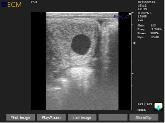
The ultrasonographic appearance of the equine embryo changes from a round shape (day 12 to 16) to a more triangular (“guitar pick”) shape on days 17 to 21. Equine embryos grow an average of 3 to 5 mm per day from day 11 to day 16 of pregnancy. Failure to grow, reduced growth rate, or failure to advance in developmental stage may be associated with eventual embryo loss. Growth plateaus between 17 and 24 days, after which growth resumes at a slightly slower rate. The developing embryonic vesicle may have a somewhat irregular outline from day 25 to 35 after the capsule has been lost.
The embryo proper may become visible 20 to 23 days after ovulation and a heart-beat can usually be observed on about day 25. If a heartbeat is not detected at day 25, the mare should be re-examined 24 to 48 hours later before a final diagnosis of embryonic loss is made and prostaglandins administered.
An additional ultrasound examination may be performed at day 35 to confirm pregnancy status as the endometrial cups are beginning to form. Mares determined to be open (not pregnant) at the day 35 examination can usually be rebred that season in the absence of endometrial cups. Administration of prostaglandins may be required to bring these mares back into heat due to persistence of the corpus luteum. Mares that are determined to be pregnant at day 35 and subsequently lose the pregnancy after day 35 will usually not cycle again that season or are very difficult to get in foal due to the presence of endometrial cups and production of equine chorionic gonadotropin.
Examination of the ovaries for the presence and characteristics of a corpus luteum and the uterus for evidence of edema are valuable during early pregnancy examinations. Maternal recognition of pregnancy occurs at approximately 12 to 14 days of pregnancy. Identification of a smaller-than-normal corpus luteum or edema in the uterus of a pregnant mare are indications that progesterone concentrations are low and that the pregnancy is at risk. The pregnancy can be maintained by initiating progesterone supplementation. Measurement of blood progesterone level may be performed to confirm the functional status of the corpus luteum.
The ovaries should be evaluated during all pregnancy examinations for the presence and physical characteristics of the corpus luteum. The primary corpus luteum should be visible and morphologically normal in a pregnant recipient mare up to day 45. Secondary or supplementary corpora lutea may be identified in pregnant mares after 45 to 60 days of gestation. Multiple secondary corpora lutea may be present on each ovary and the size of the ovary is subsequently increased. It should be noted that secondary corpora lutea do not form in every pregnant mare. Determination of the presence or absence of secondary corpora lutea may be important for management decisions, such as to whether or not to continue administration of supplemental progestins (i.e. altrenogest) to a pregnant mare.
Other Aspects of Pregnancy Diagnosis
It is of benefit to identify, measure and map the location of uterine cysts prior to breeding a mare. In some instances a uterine cyst may be confused with an embryonic vesicle. This may be crucial and potentially frustrating during an ultrasound examination for twins. Re-evaluation of the mare after an initial ultrasound examination for pregnancy may be necessary. The facts that “cysts do not grow” and “cysts do not move” within the uterus may be beneficial in differentiation of cysts from an embryonic vesicle. The equine embryo migrates throughout the uterus until day 16 or 17 and generally increases in size each day. On a rare occasion, a final diagnosis of pregnancy status may have to wait until after day 25 to determine if an embryonic vesicle, with an embryo proper and heart-beat, is present amongst a cluster of cysts.